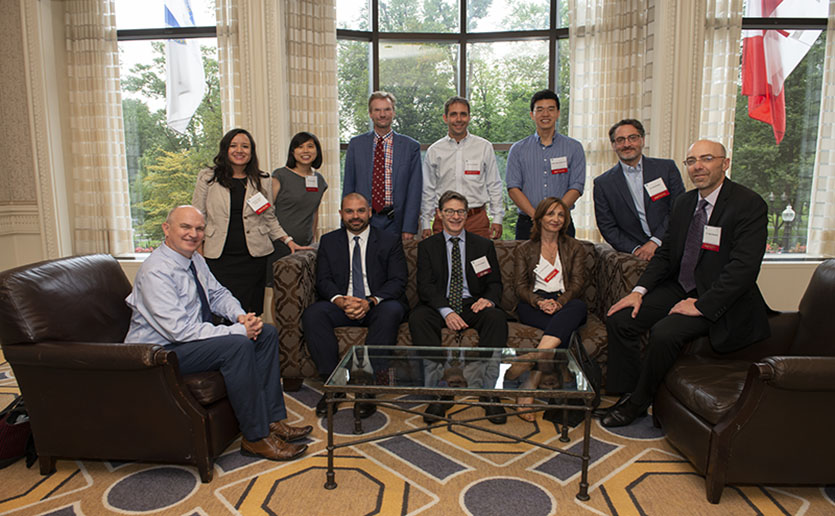
The past quarter century has seen a revolution in neuroscience. A new generation of imaging tools has yielded fundamental insights into the inner workings of the human brain, with implications for countless neurological disorders and psychiatric diseases. Many of the most important advances in this revolution have been made at the Athinoula A. Martinos Center for Biomedical Imaging at Massachusetts General Hospital — which is celebrating the 25th anniversary of its founding.
“In the past 25 years, we have come to understand better how the brain is organized and how it works,” says Bruce Rosen, MD, PhD, director of the center and vice chair for research in the Mass General Department of Radiology. “We have achieved this using technologies like functional MRI, diffusion imaging and combined PET/MR imaging — technologies that the Martinos Center pioneered here at MGH.”
The Roots of a Revolution
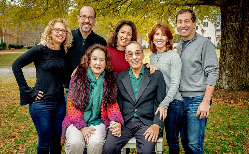
In 1999, Greek shipping magnate Thanassis Martinos and his wife Marina made a historic gift of $20 million to Massachusetts General Hospital and the Massachusetts Institute of Technology (MIT) to fund a biomedical imaging research center named for their daughter, Athinoula A. Martinos. Athinoula had passed away two years before, after struggling for years with schizophrenia. The Martinos family decided to honor her memory by supporting research that would advance our understanding of neurological disorders like schizophrenia and make progress in the development of better treatments. The Martinos Center officially launched in November 2000.
The goal of the center is to facilitate development of cutting-edge neuroimaging and other biomedical imaging technologies and to foster multidisciplinary research using those technologies to understand human disease. Their work bridges areas ranging from hardware development to the basic biosciences through to clinical investigation.
The Martinos Center was built on an existing program at Mass General focused on magnetic resonance imaging, or MRI. Led by Dr. Rosen, researchers in this earlier program had already logged major successes in pioneering neuroimaging technologies: not least, the introduction of functional MRI in 1991 and 1992, now a mainstay of several diagnostic and therapeutic interventions for a range of diseases and conditions.
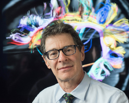
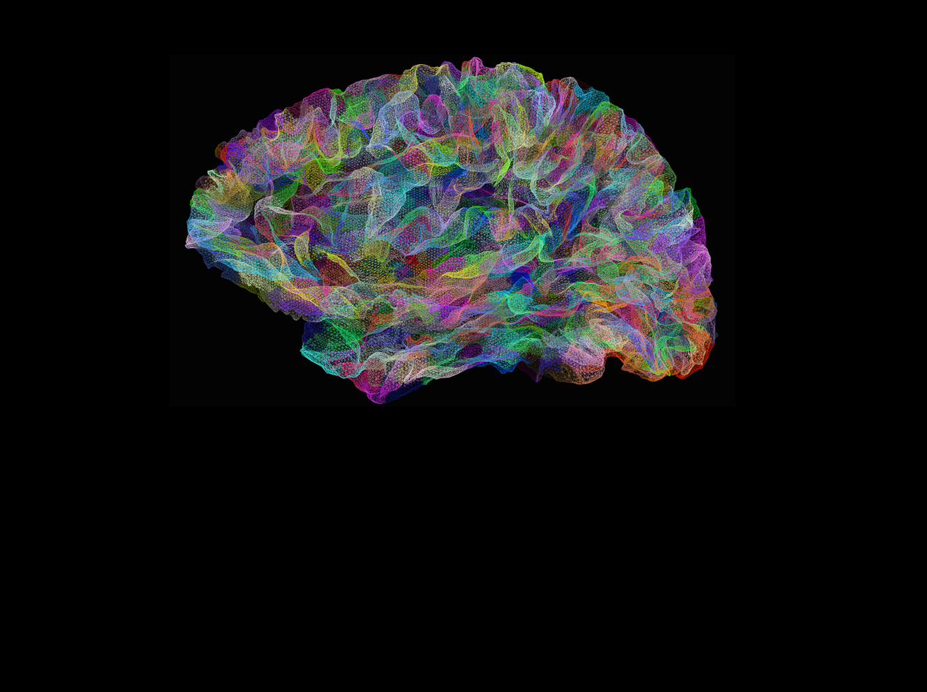
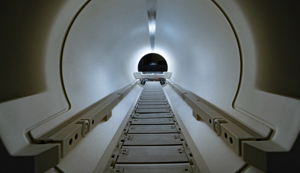
The Martinos gift enabled a major expansion of the program, nearly doubling its footprint and clearing the way for two first-of-their-kind scanners: a combined positron emission tomography (PET)/MR scanner for simultaneous measurement of brain anatomy and brain function and, later, a “Connectome” scanner capable of visualizing the connections between regions of the brain.
Advancing the Fight against Neurological Disorders
With more than 120 faculty, the Martinos Center is, today, the largest academic radiology research program in the U.S. Its contributions to improved human health include breakthroughs that are revolutionizing the diagnosis and treatment of neurological disorders and neurodegenerative diseases: Alzheimer’s disease, multiple sclerosis, autism spectrum disorder, depression and anxiety, chronic pain and many more. The wide range of projects and studies initiated by Martinos’ researchers reflects the innovative approach the Center makes possible.
Susie Huang, MD, PhD, a neuroradiologist at Mass General and associate director of the Martinos Center, uses advanced neuroimaging techniques to understand better the role of brain connectivity in neurological disorders and diseases, including multiple sclerosis among others. Currently, she leads the “Connectome 2.0” project, a groundbreaking initiative involving a state-of-the-art MRI scanner designed to map the structural connections in the brain with unprecedented detail and installed in 2023. The deep insights she and colleagues gain from this work will ultimately lead to more precise diagnoses and treatments for the neurological disorders.
Elsewhere in the center, Heidi Jacobs, PhD, is working toward earlier detection of Alzheimer’s by focusing on the locus coeruleus, a structure in the brain where a protein associated with the disease begins to accumulate in early adulthood. Imaging the amount of the protein in the locus coeruleus could help determine a person’s risk of developing Alzheimer’s earlier in the progression of the disease than is currently possible — allowing clinicians to intervene earlier to delay or even prevent the disease. Dr. Jacobs and her team have pioneered methods that make this possible and have now shown that they can use the methods to predict cognitive decline in Alzheimer’s earlier and more precisely than was previously possible.
Laura Lewis, PhD, the inaugural Athinoula A. Martinos Professor of Health Sciences and Technology at MIT and a faculty member in the Martinos Center, focuses on what the brain is doing during sleep and how this activity can contribute to neurodegenerative diseases like Alzheimer’s. She has shown, for example, that during deep sleep, large waves of cerebrospinal fluid wash over the brain, likely helping to clear waste products from the brain. This process could be crucial in maintaining brain health and developing interventions to ensure the natural process could potentially reduce the risk of Alzheimer’s and other neurodegenerative diseases.
Looking Forward
When it comes to revolutionary advances in neuro and biomedical imaging, there is no quota on innovation — and Martinos Center researchers are always looking ahead to new horizons. One of these involves molecular imaging: imaging of the living human brain as it works its biochemical magic to sustain our complex human behaviors.
“We’re moving from watching the brain’s functional anatomy — the parts of the brain are working when performing its daily tasks — to watching the actual molecular processes it undergoes during these events,” Dr. Rosen says. “We want to learn how these processes are disrupted in Alzheimer’s or schizophrenia, for example.”
Martinos Center researchers are also exploring ways to apply what they have learned about the organization of the brain to develop new types of therapy. These include neuromodulation, in which a new generation of tools will be able to stimulate or inhibit specific circuits in the brain, mitigating the effects of Parkinson’s disease, autism spectrum disorder, post-traumatic stress disorder (PTSD) and other conditions.
In addition to its work on the brain, the Martinos Center is also working on projects that image other areas of the body, expanding our understandings of various types of cancer, of liver and lung fibrosis, cardiovascular diseases, and many other diseases. In fact, the Martinos Center is planning to expand its broader capabilities in oncology with the MGB Cancer Center through the use of molecular imaging and whole-body PET-CT scanning, the first of its kind here in New England.
“The Martinos Center is one of the crown jewels of not just Mass General Brigham, but of the academic medical world,” says David F. M. Brown, MD, President, Academic Medical Centers, Mass General Brigham. “The advances and insights made possible by the Martinos Center — and through the commitment and dedication of the Martinos family — are empowering clinical discovery not only for the patients we treat, but for patients the world over.”
To learn more or support the Martinos center, contact us or make a gift.
