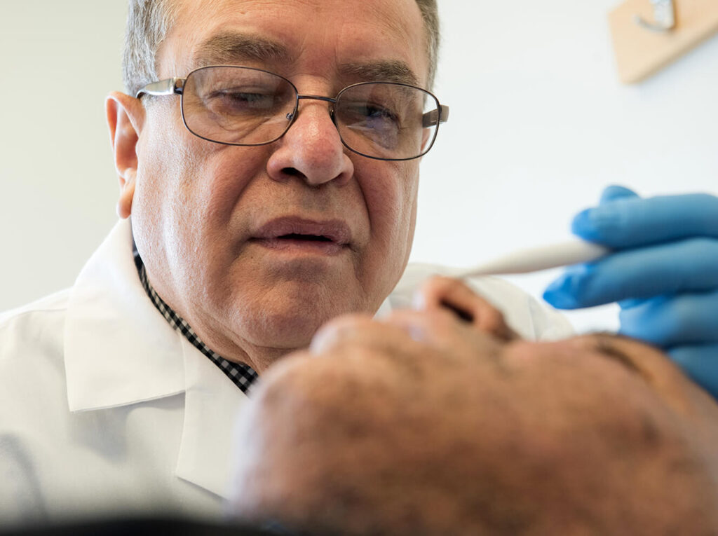
A new device that gives the human eye a “bionic edge” may sound like science fiction, says Massachusetts General Hospital dermatologist Victor Neel, MD, PhD, but it is a real and much-needed development for a procedure called Mohs surgery, a procedure to remove skin cancers on the face.

“Fred Mohs developed his technique 70 years ago, and today we’re still using the same procedure,” says Dr. Neel, director of Dermatologic Surgery at Mass General’s Department of Dermatology. “If Dr. Mohs rose from the grave, put on a white coat and went into the Mohs unit, he’d feel right at home because nothing has changed.”
Through a recent partnership with a physicist at UMass Lowell, Dr. Neel says he’s testing a device called optical polarization imaging (OPI) to allow surgeons a more accurate look at a tumor prior to surgery.
Identifying Skin Cancer Boundaries
Mohs surgeons typically use their best judgment to identify the boundary between a patient’s tumor and the normal skin around it, also referred to as the margin. “The problem,” says Dr. Neel, “is that it’s impossible to see skin cancer margins with your eyes. There’s a limitation to what the unaided eye can see.”
OPI operates like a camera by taking pictures of the collagen in the skin. Tumor cells secrete enzymes that deform and destroy collagen’s interwoven mesh-like structure. By visualizing abnormal collagen, OPI allows Dr. Neel to identify the boundary lines that separate the tumor from normal skin before making any incisions.
“It’s a very inexpensive and simple, yet elegant technology,” says Dr. Neel. “It would allow Mohs surgeons to take a picture of the surgical site in real time and construct a surgical plan based on what they are able to see.”

Successful Pilot Study
Dr. Neel emphasizes he doesn’t want to replace Mohs surgery. Instead, he says OPI could help improve accuracy and reduce the average number of surgical stages per case, saving money for patients and insurers.
Dr. Neel and his team recently published a paper showing the successful results from their first pilot study using data from six patients with basal cell carcinoma, the most common type of skin cancer. In all six cases, OPI accurately predicted the tumor’s boundaries by identifying structural collagen changes.
Dr. Neel is now in the process of detailing 60 more cases with similar results and hopes to conduct a multi-center OPI trial in the near future.
“I’m looking back at the 25,000 Mohs cases I’ve done at Mass General, and I don’t want to do another 25,000 the same way,” Dr. Neel says. “It’s exciting to see a change on the horizon.”
To learn more about how you can support the work of Mass General’s Department of Dermatology, please contact us.
A longer version of this story was first published by the Mass General Research Institute.