Magnetic resonance imaging (MRI) has become indispensable for detecting tumors, torn ligaments and other anomalies under the skin. The technology has come a long way since its introduction in 1977, and the newest machines allow radiologists to peer deep into the body with near-cellular precision. So researchers are focusing these powerful tools on the finer structures in the brain and shedding light on one of the body’s most puzzling and complex systems.
That tangle is anything but straightforward, says Dr. Yendiki: “An image of it looks like a bowl of 500,000 spaghettis.”
Anastasia Yendiki, PhD, is an assistant physicist at Massachusetts General Hospital and a researcher at Mass General’s Martinos Center for Biomedical Imaging. She studies the role of white matter. This consists of bundles of axons — threadlike extensions of nerve cells — that weave together to form the brain’s underlying connections, sometimes called the connectome. That tangle is anything but straightforward, says Dr. Yendiki: “An image of it looks like a bowl of 500,000 spaghettis.”
She and her colleagues equipped their MRIs with high-gradient coils and other enhancements, allowing them to track the random motion of tiny water molecules through the brain. Their movements reveal white matter pathways, just as tracking the movement of cars can determine where roads are, says Dr. Yendiki. She has developed new software (christened TRACULA) to help her and other researchers sort out the course and strength of brain connections.
Mapping the Brain’s Side Roads
Scientists have already mapped the brain’s primary white matter highways, says Dr. Yendiki, “but we don’t know all the smaller roads leading on and off.” Dr. Yendiki, also an assistant professor of radiology at the Harvard Medical School, is exploring these side roads — and how they vary in people with depression and other brain disorders.

The Human Connectome Project, launched in 2009, is building a database of many connectomes that could help identify additional subtypes of complex diseases. For example, if the connectome is wired differently in people with depression, that information could be used to identify subtypes of the disease based on how they manifest in brain activity rather than in patterns of behavior, which is how psychologists currently identify them. This might guide more targeted and effective treatments for bipolar disorder, chronic depression and psychotic depression.
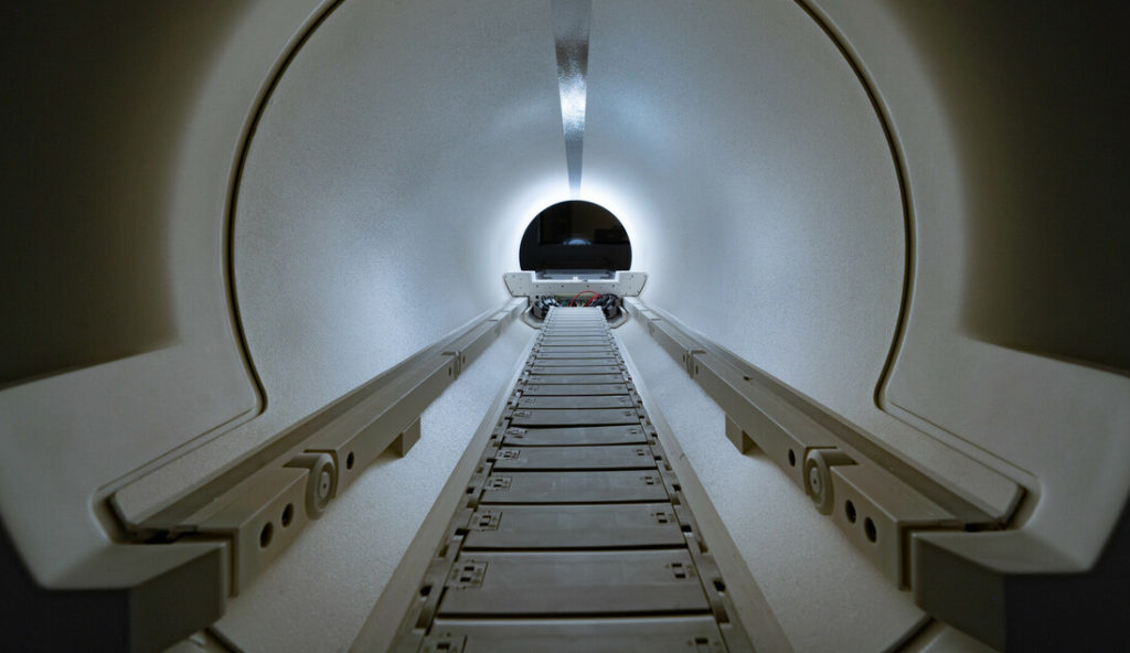
Other machines are equipped to take an even deeper dive. An MRI’s magnet causes molecules in the body to release energy that can then be recorded, decoded and finally translated into an image. A gold-standard clinical MRI uses a 3 Tesla magnet, which is about 300 times as strong as one you would put on the refrigerator door. But the newest machines, which weigh about 80,000 pounds and cost $7 million or more, use magnets capable of 7 Tesla and offer images with unparalleled resolution.
For information on how to support the research at the Martinos Center for Biomedical Imaging, please contact us.
A longer version of this article first appeared in Proto Magazine.