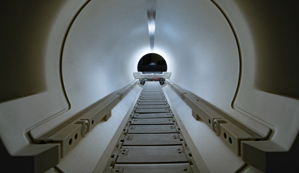
When it was first introduced in the late 1970s, magnetic resonance imaging (MRI) was a diagnostic revelation. Suddenly, doctors could see brain tumors or multiple sclerosis lesions without exposing a patient to dangerous radiation or making a single cut. But there were drawbacks. The machines were the size of U-Haul trailers, and they were expensive, which put them out of reach for many medical centers. They also relied on powerful superconducting magnets that needed to be contained in copper-shielded rooms, often located in hospital basements or offsite containment units.
Forty years later, the technology has evolved considerably. Today’s scanners are capable of capturing the tiniest structural details and tracking complex brain functions, like speech and memory, in real time. Some things, however, haven’t changed: The machines are still big, still expensive and still need to be shielded. But thanks to the work of Matt Rosen, PhD, an iconoclastic physicist in Massachusetts General Hospital’s Athinoula A. Martinos Center for Biomedical Imaging, MRI is finally ready to escape its confines.
Portable MRI
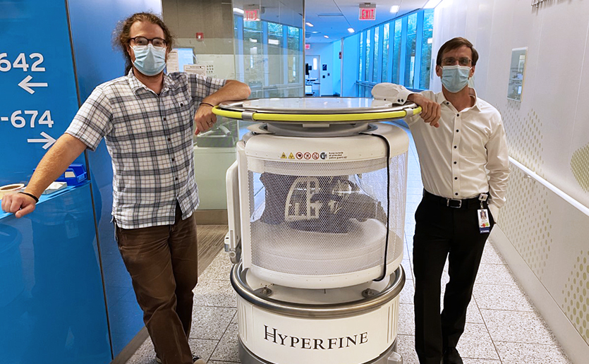
In late 2019, Connecticut-based technology firm Hyperfine Research, Inc., unveiled a new “bedside” MRI scanner inspired by Dr. Rosen’s groundbreaking work. The portable scanner is comparatively small — about the size of an upright oil drum on wheels — lightweight and able to be plugged into a standard wall socket. And while it lacks the image resolution of a standard $3 million clinical scanner, it also comes at a fraction of the cost — less than $50,000.

“Instead of having to bring the patient to the MRI, you can bring the MRI to the patient,” Dr. Rosen says. If widely adopted, Dr. Rosen’s technology could make MRI available for a variety of new clinical settings — such as the emergency room or intensive care unit — as well as for rapid-response situations, like natural disasters. It could also transform health care for the more than 4 billion people worldwide who lack access to medical imaging.
“The beauty of this system is that it doesn’t have to be the centerpiece of a giant radiology department,” says Martinos Center Director Bruce Rosen, MD, PhD (no relation to Dr. Matt Rosen). “It could be in an exam room at Mass General or in a roadside clinic in rural India.”
A Low-Powered Approach
Every water molecule in the human body carries a tiny magnetic charge. MRI scanners employ powerful magnetic fields to bring those molecules into alignment. The stronger the magnetic field, the more molecules align, the better the image resolution. But there’s a downside.
Your average top-of-the-line clinical MRI uses a 3-tesla magnet, meaning it produces a magnetic field 60,000 times more powerful than Earth’s geomagnetic field. That’s powerful enough to erase the credit cards in your wallet and turn average hand tools into deadly projectiles. But does an MRI always have to be that powerful to get results? Not according to Dr. Matt Rosen.
“Instead of having to bring the patient to the MRI, you can bring the MRI to the patient.”
In 2004, with a grant from NASA, he developed a “low-field” imaging system that generated a 6.5 millitesla (mT) field — roughly the same strength as the magnets on your refrigerator — to study how gravity affects the lungs. Later, when the U.S. Department of Defense needed a way to quickly and accurately diagnose blast-related traumatic brain injury in the field, Dr. Matt Rosen and his team capitalized on advances in physics, computer science and signal processing to create a low-field MRI scanner that was safe, effective and small enough to be easily deployed.
“Once you get rid of the big magnet, the whole concept of the machine changes,” he says.
Seeing Results

Dr. Matt Rosen co-founded Hyperfine Research with geneticist and entrepreneur Jonathan Rothberg in 2014. After six years of development, the Hyperfine portable 64 mT MRI system was approved for patient use in February 2020.
A recent study published in JAMA Neurology, by Dr. Matt Rosen and W. Taylor Kimberly, MD, PhD, chief of Neurocritical Care at Mass General, showed the portable system could safely detect stroke, brain injury and even evidence of COVID-19.
“Think of it like the cellphone,” says Dr. Bruce Rosen. “There was a time you had to be a millionaire to have one. What does MRI become when it becomes ubiquitous? I think we could see them everywhere.”
To learn about how you can support the Martinos Center, please contact us.
The Athinoula A. Martinos Center for Biomedical Imaging at Massachusetts General Hospital is one of the world’s premier research centers devoted to development and application of advanced biomedical imaging technologies. The center is part of the department of Radiology at Mass General and is affiliated with Harvard Medical School and MIT. In November 2020, the Martinos Center celebrated its 20th anniversary of creating tools to solve complex challenges in neuroscience, oncology, cardiology and more.


